Photoionization Studied With COLTRIMS

When an inner shell electron is ejected near threshold from a constituent of molecule, the outgoing photoelectron diffracts in the molecular potential. The resulting diffraction pattern reveals information on both the scattering process and the potential. By using momentum imaging techniques to measure in coincidence the dissociation fragments and the photoelectrons from the ionization of the C 1s electron in CO, we are able to obtain full images of the diffraction patterns of the CO potential as sampled by low energy electrons (up to 30 eV above threshold).


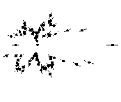
The molecular fragments are used to hold the molecules "fixed in space" relative to the polarization vector. This movie shows how the polar distribution of electrons evolves as the CO molecule is rotated relative to the polarization.

This 580x580 AVI-style clip is 1.94 MB in size. For those with smaller displays, it is also available as a 300x300 AVI (1.3 MB) or MPEG (635 kB) file.

electron-rotated.gif (3178 bytes)

 
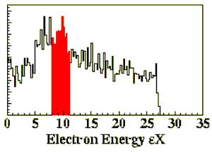
energy.gif (5900 bytes) This clip shows how the pattern at a fixed molecular orientation evolves as the photon energy is scanned.


The polarization vector of the photon is horizontal in all figures.

This 628x628 AVI-style clip is 587 kB in size. For those with smaller displays, it is also available as a 300x300 AVI (444 kB) or MPEG (428 kB) file.


The latest results and earlier work on this topic are also available.
Further information on this research is available from Professor Lew Cocke.
 
Last updated on Wednesday, 20-Jul-2005.